

Important: This article is educational and not medical advice. A routine eye exam cannot diagnose Alzheimer’s disease. Patients with medical concerns should consult their physician.
Interest in the connection between eye health and brain health has grown in recent years. Because the eye contains delicate nerve tissue linked to the brain, researchers are studying whether subtle visual changes might provide early clues about neurological conditions.
That does not mean an eye doctor diagnoses Alzheimer’s disease. Instead, optometrists may observe patterns that suggest further medical evaluation could be helpful. Understanding what an exam can and cannot reveal helps patients interpret this research responsibly.
Why Researchers Study the Eye in Alzheimer’s Disease
The retina is not merely brain-like. It develops from the same embryonic tissue as the brain and remains part of the central nervous system. Unlike the rest of that system, it can be viewed directly and noninvasively during an exam, which is exactly why researchers keep returning to it.
Research from Indiana University School of Medicine, published in Alzheimer’s & Dementia, found that a gene variant known to raise Alzheimer’s risk in humans impairs retinal function in mouse models. The authors describe retinal imaging as a promising noninvasive way to detect early neural changes, though the work has not yet been replicated in patients.
The Retina as Neural Tissue
The retina contains nerve cells that communicate directly with the brain. Because Alzheimer’s affects nerve pathways, researchers are investigating whether similar changes occur in the eye. These changes are not visible to patients but can sometimes be detected through imaging.
Blood Vessel and Structural Changes
The retina’s microvasculature closely mirrors that of the brain. Researchers are studying whether vessel narrowing or reduced density tracks are associated with early neurological decline. The work is promising but not yet conclusive.
Why Imaging Technology Matters
Modern eye imaging allows doctors to document microscopic structures over time. This capability makes the eye a useful location for studying disease progression without invasive testing.
What a Routine Eye Exam Can Actually Observe
A routine eye exam evaluates more than vision clarity. It examines structures that may reflect broader health patterns.
1. Structural Retinal Changes: Optical coherence tomography maps the retina layer by layer at micron-level detail, letting your optometrist compare today’s scan against years of prior visits. Measurable thinning may prompt further evaluation.
2. Vascular Patterns: Blood vessel health can reflect systemic conditions. Eye doctors often observe vascular patterns that suggest medical follow-up.
3. Functional Vision Changes: Shifts in contrast sensitivity or color discrimination can appear before patients notice any loss of clarity. These findings alone are not diagnostic but contribute to the clinical context.
These observations provide context rather than conclusions. Each falls within the wider set of signs an optometrist screens for at every visit, and when a pattern looks unusual, it helps determine whether further medical evaluation is warranted.
What an Eye Exam Cannot Diagnose
Understanding the limits of an eye exam is just as important as understanding its benefits. While modern imaging can reveal structural changes in the eye, these findings alone cannot determine the presence of Alzheimer’s disease.
1. It Cannot Confirm Alzheimer’s Disease
An eye exam does not evaluate memory, cognition, or brain function. Even if retinal changes are observed, they are only considered indicators that further medical testing may be helpful. Moreover, optometrists identify visual and structural observations, not cognitive disorders. The findings may raise awareness, but do not establish disease.
2. It Cannot Replace Neurological Testing
Diagnosis requires cognitive testing, brain imaging, and physician assessment. Retinal findings carry meaning only when interpreted alongside that broader picture, never on their own.
3. It Cannot Predict Future Cognitive Decline
Retinal patterns cannot determine whether a person will develop dementia. They only contribute to a larger clinical picture evaluated by medical specialists. When unusual patterns appear, your optometrist coordinates with your primary care physician or a specialist. The goal is early communication, not independent diagnosis, eye care that supports your medical team rather than substituting for it
How Art of Optiks Supports Early Awareness
Preventive care works best when providers collaborate. Eye exams can contribute valuable information within a broader healthcare plan.
1. Baseline Monitoring – Regular exams create a record of normal findings, which matters because vision shifts naturally with age, and normal change has to be distinguished from concerning change.
2. Documenting Changes – Small variations can be tracked and communicated to physicians if needed. This supports earlier medical conversations.
3. Coordinated Care – Optometrists help patients understand whether additional evaluation may be beneficial, whether that means a physician referral or ongoing monitoring of an eye condition.
By tracking subtle changes over time, eye doctors contribute useful information that may support earlier medical conversations. This collaborative approach helps ensure patients are guided appropriately without replacing physician evaluation.
Why Preventive Eye Exams Matter Beyond Vision
Routine eye care evaluates sight and ocular health together. The retina is one of the few places a clinician can view blood vessels and nerve tissue directly, which is why diabetes, hypertension, and high cholesterol are sometimes flagged first at an eye exam rather than a physical.
Through comprehensive evaluations at Art of Optiks, patients build a documented record of change over time. The risks that record guards against shift by decade, the eye conditions that matter at 40 are not the ones that matter at 70, and each is easier to catch against a baseline than in isolation.
Understand Your Eye Health With a Professional Evaluation
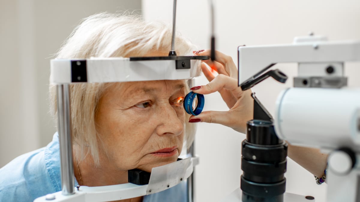
A routine eye exam cannot diagnose Alzheimer’s disease, but it can contribute to early awareness by documenting subtle changes over time. Understanding this distinction helps patients use preventive care appropriately and avoid misinterpreting research headlines.
If you are due for an exam or want to establish a baseline, Contact Art of Optiks today. Regular exams support both your vision and your broader health awareness, so you know when a conversation with your physician is worth having